Retinal vein occlusion
Retinal vein occlusion overview
Retina is a delicate tissue in eye that needs the flow of blood from arteries & veins. Any clots or blockage or occlusion of a vein leads to retinal vein occlusion.
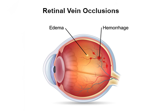
The main reason for this occlusion or blockage is one vessel pressing another, which is resulting in narrowing and blocking of vessel. In some cases, an occlusion can occur even due to blood clot. RVO can result in fluid leakage from the blood vessels, which tends to swelling and thickening of retina. The inflammation of retina is known as macular edema—which causes blurry vision or vision loss.
Another risk with Retinal vein occlusion, is the unusual growth of new blood vessels either at the backside or front side of the eye. If new blood vessels grows at the backside of the eye, then it causes bleeding. On the other hand, if new blood vessels grows at the front side of the eye, it causes pressure in eye to go up and too much pressure can damage the vision permanently. Before it gets too late, get in touch with our retinal specialists for Retinal vein occlusion treatment.

Types of Retinal vein occlusion
There are two types of Retinal vein occlusion namely, BRVO (Branch Retinal Vein Occlusion) and CRVO (Central Retinal Vein Occlusion).
Retinal vein occlusion causes
Retinal vein occlusion is caused due to arteries hardening & blood clots. In people with narrowed or damaged blood vessels, blockages are common. Even in people with few chronic conditions blood clots are often seen. The following are some of the chronic conditions and risk factors that increase the risk of Retinal vein occlusion:
- Diabetes
- Glaucoma (increased eye pressure)
- Blood vessel diseases or inflammations (vascular disease)
- High blood pressure
Retinal vein occlusion Symptoms
Below are the some of the symptom of Retinal vein occlusion:
- Blurriness or loss of vision
- Eye pain
- Floaters
If you are experiencing any of the above symptoms, get evaluated by our Challa Eye Care Centre retina specialists right away.
How Retinal vein occlusion is diagnosed?
Fluorescein Angiography and Optical coherence tomography tests are often used to identify the extent of harm from a retinal vein occlusion.
In Fluorescein Angiography, A yellow dye known as fluorescein is inserted into vein in the arm, then a camera is used to click special pictures of inside of the eye to look at the blood flow in the retina.
In Optical coherence tomography, retina is scanned and detailed images are provided to identify swelling & leakage of fluid in retina.
Retinal vein occlusion treatment at Challa Eye Care Centre
Our experts at Challa Eye Care Centre evaluate the extent of and damage to the eye caused by retinal vein occlusion and teat appropriately by using lasers and intravitreal injections or both.